Papers by Roger Schmidt Brock
Revista da Associação Médica Brasileira, 2019
Arquivos Brasileiros de Neurocirurgia: Brazilian Neurosurgery, 2020
Neurological Sciences, 2019
Purpose Symptomatic Chiari type I malformation (CM) is treated with posterior fossa decompression... more Purpose Symptomatic Chiari type I malformation (CM) is treated with posterior fossa decompression with/ without duroplasty. Few authors suggested cerebellar tonsil caudal migration due to a supposed "caudal traction" of cranial nerve structures in a socalled occult tethered cord syndrome. For these authors, filum terminale (FT) sectioning may improve CM symptoms. The objective of this review is to evaluate the effect of FT sectioning on the treatment of CM. Methods Using the PRISMA guidelines for systematic reviews, we reviewed studies to evaluate patient's outcomes with CM who underwent FT sectioning. The MINORS instrument was used for methodological quality assessment. The included studies' levels of evidence (LOE) were classified according to the Oxford Centre of Evidence-Based Medicine. Results Two studies from the same group of authors were included. We cannot assure if the cited cases in the first study were also included in their latter published study. The described results suggest that outcomes were not collected in a standardized fashion. Outcomes are described vaguely as a percentage of improvement. Case series samples were small and included not only patients with CM but also patients with scoliosis and syringomyelia. The MINORS score reported that both studies had low methodological quality. Both included studies were classified as level 4 of evidence.
XXXII Congresso Brasileiro de Neurocirurgia, 2018
The Chiari Malformations, 2020
The advent of intraoperative ultrasonography (USG) has allowed identification of craniocervical j... more The advent of intraoperative ultrasonography (USG) has allowed identification of craniocervical junction (CVJ) anatomy and cerebrospinal fluid dynamics and CVJ structures with real-time images. It is possible to use intraoperative USG in patients with Chiari malformation type I as a method for selection of candidates for posterior fossa decompression with bone removal alone. This chapter describes the authors’ experience using intraoperative USG.
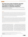
Spinal cord epidural metastases (SEM) are a common complication of systemic cancer, and has an in... more Spinal cord epidural metastases (SEM) are a common complication of systemic cancer, and has an increasing incidence in oncological patients 1. From the vertebral body, these metastases can invade into the vertebral canal and cause spinal cord compression and vascular insufficiency, leading to myelopathy, cauda equina, or root dysfunction syndromes. Pain is the most common symptom, present in 83-95% of these patients 1. Also, two thirds of them have motor signs at the time of diagnosis and sensory deficits can be detectable in their majority 2. Bowel and bladder disturbances tend to appear later, but about half of patients with SEM are already catheter dependent at diagnosis 1. Unfortunately, SEM is part of an already spread cancer disease and its treatment should focus on improving the patient's quality of life 3. Several protocols and strategies about how to treat patients with SEM, including surgery, radiotherapy and chemotherapy have been studied but the debate is still open on which are the patients who will benefit more from each treatment. Surgical treatment can lead to accurate diagnosis, control refractory pain to non-operative measures, preserve or restore neurological function, and maintain alignment and stability of the bony spine 4 , but it is also accepted that patients with limited prognosis should not be candidates
Revista da Associação Médica Brasileira
This guideline aims to analyze the effect of the section of the filum terminale in the treatment ... more This guideline aims to analyze the effect of the section of the filum terminale in the treatment of Chiari malformation type I symptoms. CONFLICT OF INTEREST There is no conflict of interest related to this review that can be declared by any of the authors. * the maximum miNorS score for non-randomized studies is 16 points. therefore, the methodological quality of both studies selected is low.
Journal of Clinical Neuroscience
Lista de Gráficos Gráfico 1 Distribuição dos pacientes quanto a presença de velocidade de fluxo d... more Lista de Gráficos Gráfico 1 Distribuição dos pacientes quanto a presença de velocidade de fluxo de líquido cefalorraquidiano acima de

Arquivos Brasileiros de Neurocirurgia: Brazilian Neurosurgery
The surgical treatment of intracranial aneurysms is a routine operation in the neurosurgeon pract... more The surgical treatment of intracranial aneurysms is a routine operation in the neurosurgeon practice. Complex aneurysms are those with morphological irregularities, usually large or giant; thrombosed, partially thrombosed or calcified; with aberrant fundus/neck ratio and near eloquent neurological structures. These cases demand special skills by the surgical team. The present article is a report of two cases of complex aneurysms successfully treated, with a discussion on the role of neurophysiological monitoring. In these two cases of supra- and infratentorial complex giant aneurysms, intraoperative monitoring was extremely relevant. Thus, we believe that treating complex and giant aneurysms carries several pitfalls, and the use of multimodal intraoperative monitoring is mandatory to mitigate risks and deliver the best result to the patient.

Surgical Neurology International
Background: Hemangioblastomas account for about 1%–3% of all central nervous system tumors. They ... more Background: Hemangioblastomas account for about 1%–3% of all central nervous system tumors. They are usually associated with the Von Hippel–Lindau syndrome and typically occur in the posterior fossa, or throughout the spinal neuraxis. Here, we report the unusual case of a sporadic cauda equina hemangioblastoma. Case Description: A 28-year-old Caucasian female patient presented with progressive low back pain of 2 months duration. The magnetic resonance (MR) revealed a heterogeneous intradural and extramedullary lesion at the L2 level; with intravenous contrast, there were vascular flow voids and surrounding vasogenic edema (i.e., measuring 4.1 cm × 3.5 cm). The patient underwent an L2 right hemilaminectomy under intraoperative neurophysiological monitoring. She was discharged the 4th postoperative day, neurologically intact. Literature describes 21 previous reports of sporadic isolated spinal hemangioblastomas. Conclusion: Although rare, sporadic, and isolated hemangioblastomas of th...

JBNC - JORNAL BRASILEIRO DE NEUROCIRURGIA
Introduction. Surgical posterior fossa decompression of Chiari malformation type 1 (CM-I) is reco... more Introduction. Surgical posterior fossa decompression of Chiari malformation type 1 (CM-I) is recommended in symptomatic patients. The classic surgery is a suboccipital craniectomy and C-1 laminectomy, with duroplasty. However, a range of complications associated with surgical management of CM-I includes pseudomeningocele, CSF leakage, aseptic meningitis, and wound infections, making dural closure and reconstruction one important step to determine morbidity. Our objective is to present a technique of pericranium harvest and dural closure and describe our experience. Methods. A retrospective study was conducted based on records of patients treated in Hospital das Clínicas of the University of São Paulo, diagnosed with CM-I and submitted to posterior fossa decompression from January 2008 until May 2015. We evaluated the occurrence of post-operative complications of symptomatic pseudomeningocele or incisional CSF leak. The occurrence of meningitis, surgical site infection or other compl...
JBNC - JORNAL BRASILEIRO DE NEUROCIRURGIA
Background: Primary brain tumors associated with cerebral arteriovenous malformations (AVM) altho... more Background: Primary brain tumors associated with cerebral arteriovenous malformations (AVM) although known is a rarely reported finding. There have been approximately 50 cases reported. Only two cases of a single lesion with coexistence of AVM and glioblastoma were described. Case Report. We report a case of a 46-year-old woman with headache and seizures for 2 months who performed a MR which displayed a large right frontal lesion suggesting a glioblastoma. Results. During surgical resection, two large veins could be seen draining tumoral vascularization in the surrounding cortex. After reaching the deepest portion of tumor, veins were coagulated and cut to allow tumor removal. However, unexpected bleeding started to occur with associated lesion growth and edema, resembling an AVM. Material sent to pathology revealed giant glioblastoma in association to an AVM with some thrombosed vessels and ischemic necrosis. Two previous studies discussed similar cases. Conclusion. The need of mag...
Arquivos Brasileiros de Neurocirurgia: Brazilian Neurosurgery
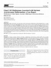
Cisternal spinal accessory schwannoma are still a rare condition without neurofibromatosis with o... more Cisternal spinal accessory schwannoma are still a rare condition without neurofibromatosis with only 32 cases reported so far. We describe a cisternal accessory schwannoma presented in a 36-year-old woman with posterior cervical pain and cervical mieolopaty, defined by grade IV tetraparesia. A suboccipital craniectomy with C1 posterior arch resection was performed. During microsurgical dissection together with electrophysiological monitoring and nerve stimulation tumor was identified as having the spinal accessory root as its origins. Carefully intraneural dissection was then performed with complete lesion removal, histopatological examination confirmed the hypothesis of schwannoma. The patient was free from pain and improved her neurological status with no accessory nerve palsy. Complete surgical resection is indicated for such lesions and can be achieved with good outcome.

Arquivos Brasileiros de Neurocirurgia: Brazilian Neurosurgery
ResumoDescrevemos os resultados laboratoriais de um sistema de transdução modificado que elimina ... more ResumoDescrevemos os resultados laboratoriais de um sistema de transdução modificado que elimina a maioria das desvantagens de sistemas hidrostáticos convencionais mencionados na literatura.Foram calculados medidas de acurácia absoluta, desvio-24 horas e testes dinâmicos de pressão. O ajuste da regressão linear utilizando-se os valores médios das 10 medidas efetuadas para cada transdutor propiciou coeficientes de correlação significativos. Os testes de desvio-24 horas foram muito semelhantes entre os transdutores comparados, não havendo diferença estatística. O teste dinâmico também foi extremamente significativo: coeficiente de correlacão (r) = 0,9999; (sd) = 0,78; p < 0,0001.Concluiu-se que a modificação do sistema reproduziu valores tão corretos quanto o sistema tradicional; o sistema hidrostático modificado pode se constituir em uma boa alternativa técnica neurocirúrgica se a sua funcionalidade for comprovada na prática.

Pediatric neurosurgery, 2018
A 4-year-old girl was admitted to the emergency department after having been buried beneath a wal... more A 4-year-old girl was admitted to the emergency department after having been buried beneath a wall. A computed tomography scan revealed anterior grade V L5-S1 spondylolisthesis, and magnetic resonance imaging showed a traumatic rupture of the fibrous annulus of the L5-S1 intervertebral disc and lesion of the anterior longitudinal and yellow ligaments. The patient underwent anterior and posterior fixation. Four months later she was able to walk independently, despite a persistent left foot drop. Additionally, we conducted a literature review on lumbosacral spondyloptosis in the pediatric population published between 1990 and 2017. We found 16 cases, 86.6% of which were male, with a mean patient age of 16 ± 5.05 years. Most patients underwent spine instrumentation. Based on the data reviewed, the neurological status at admission might be a valid predictor of outcome. Pedicle screws are a safe and reliable procedure for stable fixation of the spine in these cases. The removal of screws...
World neurosurgery, Jan 15, 2017
Halo fixation is one of the possible treatments for cervical spine fractures. However, improper u... more Halo fixation is one of the possible treatments for cervical spine fractures. However, improper use of these devices may lead to many complications, such as pin loosening, halo dislocation, pin site infection, and intradural penetration. We report the case of a 43-year-old man who first presented with a seizure and an altered level of consciousness 5 months after halo-vest placement for an odontoid fracture. Brain imaging showed a brain abscess, under the previous left parietal pin. The patient underwent abscess drainage and antibiotics were administered for 12 weeks. On hospital discharge, he presented with only mild impairments. Misapplication of halo fixation devices may lead to serious complications, including intracranial pin penetration and brain abscesses. Proper use of the recommended technique may decrease the risk for complications related to the procedure.

Arquivos de Neuro-Psiquiatria, 2017
In the present study, we evaluated the reliability and safety of a new upper cervical spine injur... more In the present study, we evaluated the reliability and safety of a new upper cervical spine injury treatment algorithm to help in the selection of the best treatment modality for these injuries. Methods Thirty cases, previously treated according to the new algorithm, were presented to four spine surgeons who were questioned about their personal suggestion for treatment, and the treatment suggested according to the application of the algorithm. After four weeks, the same questions were asked again to evaluate reliability (intra- and inter-observer) using the Kappa index. Results The reliability of the treatment suggested by applying the algorithm was superior to the reliability of the surgeons’ personal suggestion for treatment. When applying the upper cervical spine injury treatment algorithm, an agreement with the treatment actually performed was obtained in more than 89% of the cases. Conclusion The system is safe and reliable for treating traumatic upper cervical spine injuries. ...
Uploads
Papers by Roger Schmidt Brock