Papers by Masahiko Nakasono
European journal of clinical investigation, Apr 18, 2024
Journal of Gastroenterology and Hepatology, Jan 10, 2024
Gastric hamartomatous inverted polyp (g-HIP) is rare gastric elevated lesion forming endophytic g... more Gastric hamartomatous inverted polyp (g-HIP) is rare gastric elevated lesion forming endophytic growth pattern which etiology remains unknown. G-HIP is said to be associated with gastric cancer and gastritis. We systematically reviewed Japanese g-HIP’s clinicopathological features reported before2002. Japanese g-HIP amount to20lesions,18patients ; 7(38.9%)were males and 11(61.1%)were females. The mean age of patients was 60.2 years old. The mean size of the lesions was21.7millimeter. No of the polyp shape was Yamada type IV7, Yamada type I/flat elevation 6, Yamada type III 4 and Yamada II 3, respectively. The site of the lesions was Body11, Cardia3, Fornix3and Antrum1, respectively. In12described cases, accompanied lesion was Gastritis, Carcinoma, GCP, Hyperplastic polyp, GIST and remnant stomach due to ulcer. Of the submucosal shaped(Yamada type I/flat elevation)g-HIP,50% has GCP and62.5% has gastric cancer. The common feature of g-HIP was pyloric gland-like mucous gland proliferat...
Scientific Reports, Sep 26, 2022
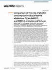
The clinical difference between nonalcoholic fatty liver disease (NAFLD) and metabolic-associated... more The clinical difference between nonalcoholic fatty liver disease (NAFLD) and metabolic-associated fatty liver disease (MAFLD) between the two sexes is unclear. This study aimed to determine the influences of alcohol consumption and qualitative abdominal fat between male and female patients with NAFLD and MAFLD. This cross-sectional study examined 11,766 participants who underwent health checkups comparing lifestyle habits, biochemical features, and noninvasive liver fibrosis scores, between non-MAFLD and MAFLD groups. Furthermore, differences in alcohol consumption and qualitative abdominal fat were examined between male and female patients with NAFLD and MAFLD. The prevalence of metabolic dysregulation, ratio of visceral fat area to subcutaneous fat area, and noninvasive liver fibrosis scores were significantly higher in male patients with MAFLD than in those with NAFLD (p < 0.05), but these were not significantly different in female patients. Among male patients with an alcohol consumption of > 70 g/week, several noninvasive liver fibrosis scores were significantly higher in the MAFLD group than in the NAFLD group (all p < 0.05). The influences of alcohol consumption and qualitative abdominal fat on NAFLD and MAFLD were different between sexes. The development of liver fibrosis should be considered in male patients with MAFLD who exceed mild drinking. Nonalcoholic fatty liver disease (NAFLD) is currently the most common liver disease in Asian and Western countries, and it may lead to nonalcoholic steatohepatitis, cirrhosis, liver failure, and hepatocellular carcinoma 1-5. NAFLD is diagnosed by the presence of hepatic steatosis in the absence of excessive alcohol consumption or other liver diseases, and it is known to be strongly associated with metabolic syndrome 6,7. However, the presence of metabolic dysregulation has not been used in the definition of NAFLD. Recently, metabolic-associated fatty liver disease (MAFLD) was proposed from an international expert consensus in 2020 8 , which highlights the association between fatty liver disease and metabolic dysregulation and does not require the exclusion of excessive alcohol consumption, viral hepatitis, or other liver diseases 8,9. Although there are many reports on the influences of alcohol consumption and abdominal fat on NAFLD 10-14 , the utility of MAFLD in clinical practice and the influences of alcohol consumption, qualitative abdominal fat, and sex when distinguishing between MAFLD and NAFLD are not sufficiently clear because the criteria for MAFLD are new and do not assess alcohol consumption and qualitative abdominal fat. Therefore, this study aimed to investigate the clinical factors associated with MAFLD (including MAFLD subgroups) to clarify the clinical differences between NAFLD and MAFLD based on alcohol consumption, qualitative abdominal fat, and sex.
Clinical Nutrition, May 1, 2023
Japanese Journal of National Medical Services, 1995
Nutrients
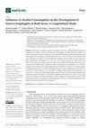
The influence of changes in alcohol consumption on erosive esophagitis (EE) development in both s... more The influence of changes in alcohol consumption on erosive esophagitis (EE) development in both sexes is unclear. This observational study investigated sex differences in the influence of alcohol consumption on EE development, and included 2582 patients without EE at baseline from 13,448 patients who underwent >2 health check-ups over >1 year. The rates of non-drinkers who started drinking, and drinkers who abstained from drinking, who increased, and who decreased their weekly alcohol consumption were 7.2%, 9.7%, 14.7%, and 24.1% and 7.3%, 17.8%, 12.8%, and 39.0% in men and women, respectively. In the final cohort, 211/1405 (15.0%) men and 79/1177 (6.7%) women newly developed EE. The odds ratio (OR) for drinking in EE development was 1.252 (95% confidence interval (CI), 0.907–1.726) among men and 1.078 (95% CI, 0.666–1.747) among women. Among men aged <50 years, the OR for drinking ≥70 g/week in EE development was 2.825 (95% CI, 1.427–5.592), whereas among women, the OR for...
Japanese Journal of National Medical Services, 1995
The Japanese journal of gastro-enterology, 2007
Humoral hypercalcemia of malignancy (HHM) in neoplastic syndrome has been most commonly reported ... more Humoral hypercalcemia of malignancy (HHM) in neoplastic syndrome has been most commonly reported in squamous cell carcinoma. Gallbladder carcinoma with HHM is uncommon. In this report, we describe a male case of gallbladder carcinoma with marked hypercalcemia and a high level of serum parathyroid hormone-related peptide (PTHrP). An immunohistochemical examination using PTHrP was also positive.
Acta Gastro-enterologica Belgica, 2004
Nihon Rinsho Geka Gakkai Zasshi (Journal of Japan Surgical Association), 2013
Gastroenterological Endoscopy, 2000
Additional file 1: Figure 1. This study protocol. BP, blood pressure; EGD, esophagogastroduodenos... more Additional file 1: Figure 1. This study protocol. BP, blood pressure; EGD, esophagogastroduodenoscopy; HF, high-frequency; HRV, heart rate variability; LF, low-frequency; POMS, profile of mood states; PR, pulse rate.
Artificial Organs, 2020
This is an open access article under the terms of the Creat ive Commo ns Attri bution License, wh... more This is an open access article under the terms of the Creat ive Commo ns Attri bution License, which permits use, distribution and reproduction in any medium, provided the original work is properly cited.
The journal of medical investigation : JMI, 2001
Congenital absence of the gallbladder is rare among biliary abnormalities, and its preoperative d... more Congenital absence of the gallbladder is rare among biliary abnormalities, and its preoperative diagnosis has been considered very difficult. We encountered a patient with congenital absence of the gallbladder and suggest a possible preoperative diagnosis of the abnormality, as well as reviewing the literature.
The Journal of Medical Investigation, 2007
Gabexate mesilate (GM) relaxes the papilla of Vater in addition to inhibiting the several proteas... more Gabexate mesilate (GM) relaxes the papilla of Vater in addition to inhibiting the several proteases. We evaluated whether prophylactic administration of GM would prevent the occurrence of acute pancreatitis in endoscopic papillary balloon dilation (EPBD). Nineteen patients with common bile duct stones were separated into two groups according to the admission year. The group A has been administered GM intravenously at 2mg/kg/hr after EPBD till six hours later, and the group B has been administered GM before fifteen minutes of EPBD till six hours later. The mean value of sphincter of Oddi (SO) basal and peak pressure in the group B was significantly lower than that in the group A, moreover the mean value of the pancreatic pressure in the group B was significantly lower than that in the group A. However two cases had mild acute pancreatitis in the group B. GM loosened SO and pancreatic duct pressure by direct stimulation of SO, although it could not have enough effect to prevent the acute pancreatitis in EPBD.

Journal of Gastroenterology and Hepatology, 2006
Colonic pseudolipomatosis is rare and the pathogenesis is controversial. The purpose of the prese... more Colonic pseudolipomatosis is rare and the pathogenesis is controversial. The purpose of the present paper was to clarify endoscopic and histological characteristics of colonic pseudolipomatosis and to discuss the etiology. A total of 15 lesions from 14 patients was reviewed. They were able to be histologically classified into two groups on the basis of variety in size of the vacuoles: Group A, the ratio of largest vacuole to smallest vacuole in size is less than three, Group B, the ratio is more than four. Four of 15 lesions were group A, and were endoscopically polypoid or flat lesions covered with normal-looking mucosa. They were microscopically characterized by (i) predominant location in the upper portion of the lamina propria; (ii) no submucosal involvement; (iii) less variation in vacuolar size; and (iv) no association with lymph follicles. The vacuoles of group A contained proteinaceous materials in two of four lesions. Group B (11 lesions) had small elevated mucosa with normal-looking surface or non-elevated reddish mucosa. Microscopically, the lesions were mainly located in the lower portion of the lamina propria, occasionally also in the submucosa, had variable-sized vacuoles, and were related to lymph follicles. It is suggested that the vacuoles in group A contain fluid, and may indicate an abnormal stagnation of interstitial fluid. Microscopic appearance of group B is essentially similar to that of pneumatosis coli. It is thought that group B probably results from penetration of gas from the crypts into the mucosa during colonoscopy. It is unclear why group B had a preference for ileocecal valve and an association with lymph follicles.
Journal of Gastroenterology and Hepatology, 2006
Duodenal lymphangitis carcinomatosa has been sporadically described, but so far little attention ... more Duodenal lymphangitis carcinomatosa has been sporadically described, but so far little attention has been paid to duodenal lymphangitis carcinomatosa. Four cases with duodenal lymphangitis carcinomatosa were endoscopically and histologically examined. The four cases exhibited multiple polypoid lesions along the Kerckring&amp;amp;amp;amp;amp;amp;amp;amp;amp;amp;amp;amp;amp;amp;amp;amp;amp;amp;amp;amp;amp;amp;amp;amp;amp;amp;#39;s folds and/or were covered by characteristically granular, non-ulcerated mucosa upon thickening. The granularity seems to been caused by dilated lymph vessels containing the carcinoma cells. The lesions were microscopically characterized by: (i) involvement of lymph vessels located in the upper portion of the lamina propria; (ii) no inflammatory changes; and (iii) no desmoplastic changes. Primary sites were thought to be the stomach in case 1, the pancreas in cases 2 and 4, and unknown in case 3. All patients died within 6 months after admission or endoscopic examination. As duodenal lymphangitis carcinomatosis shows characteristic endoscopic appearance, endoscopic diagnosis is not difficult. We should realize that the lesion represents extremely poor prognosis, and it should be distinguished from ordinary metastatic duodenal carcinoma.
Journal of Gastroenterology and Hepatology, 2007
The aim of this study was to clarify the etiology and clinical significance of solitary and scatt... more The aim of this study was to clarify the etiology and clinical significance of solitary and scattered esophageal varices by evaluating their hemodynamics and other characteristics using infrared endoscopy and endoscopic ultrasonography. The study group comprised 44 lesions of these two related types detected in 28 patients by visible-light endoscopy. Infrared endoscopy was used to characterize blue-black coloration before and after rapid intravenous injection of indocyanine green (2 mg/kg). During endoscopic ultrasonography, depth within the esophagus and echo patterns of these varices were characterized. Diameters of these varices were significantly smaller in lesions more strongly staining by infrared endoscopy. Lesion diameter was significantly smaller in varices showing homogeneous low echogenicity than in those showing mixed echogenicity. Lesions showing homogeneous high echogenicity stained most weakly followed in turn by lesions with mixed echogenicity and finally those showing homogeneous low echogenicity. Indocyanine green injection was useful for infrared observation of the hemodynamics of solitary and scattered esophageal varices, as was endoscopic ultrasonography in defining the location and morphology of these lesions. Varices with larger diameters stained more persistently when hemodynamics were evaluated by infrared endoscopy, and often showed a mixture of low and high echogenicity by endoscopic ultrasonography. These observations suggest that blood flow in the varices is slowed, and that the risk of hemorrhage increases with increased diameter especially with uniform enhancement and uniform echogenicity.


Uploads
Papers by Masahiko Nakasono