Papers by Gurudutta Gangenahalli
ACS Biomaterials Science & Engineering, Feb 24, 2023
Gene Expression Patterns, Sep 1, 2020
Differentiation of the HSCs into myeloid lineage is primarily monitored by transcription factor P... more Differentiation of the HSCs into myeloid lineage is primarily monitored by transcription factor PU.1. GATA1 acts as a negative regulator by antagonizing the function of PU.1 by bindings its β3/β4 domain. In this study, a mutation was induced in PU.1 which blocks its interaction with GATA1. The pure form of this mutant protein i.e Y244D was loaded on poly (lactic-co-glycolic acid) nanoparticles to transfect CD34 þ cells, which act as a selective tool to enhance the myelopoiesis, as confirmed by flow cytometry analysis. Further, microarray data analysis was performed to gather information on myelopoiesis specific signaling pathways and genes involved in myelopoiesis like CCL2, S100A8, and S100A9, which were also found to be significantly upregulated in the mutant form. Different molecular functions like antioxidant activity, signal transduction, developmental processes, and biological adhesion were analyzed. This study potentially signifies that PU.1 mutant is highly efficient in myelopoiesis.
Frontiers in Genetics, Jun 13, 2019
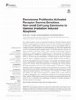
The nuclear receptors known as peroxisome proliferator activated receptor gamma (PPARG) are lipid... more The nuclear receptors known as peroxisome proliferator activated receptor gamma (PPARG) are lipid-activated transcription factors that have emerged as key regulators of inflammation. PPARG ligands have been shown to have an anti-proliferative effect on a variety of cancers. These ligands can induce apoptosis via TP53 (Tumor protein p53) or ERK1/2 (Extracellular signal-regulated kinases 1/2) (EPHB2) pathways. However, the exact mechanism is not known. PPAR, a type II nuclear hormone receptor deserves attention as a selective target for radiotherapy. Our study examines the potential of selective agonism of PPARG for radiation therapy in non-small cell lung carcinoma (NSCLC). We found that the overexpression of PPARG protein as well as its induction using the agonist, rosiglitazone was able to stimulate radiation-induced cell death in otherwise radio resistant NSCLC A549 cell line. This cell death was apoptotic and was found to be BAX (BCL2 associated X) mediated. The treatment also inhibited radiationinduced AKT (Protein Kinase B) phosphorylation. Interestingly, the ionising radiation (IR) induced apoptosis was found to be inversely related to TP53 levels. A relatively significant increase in the levels of radiation induced apoptosis was observed in H1299 cells (TP53 null) under PPARG overexpression condition further supporting the inverse relationship between apoptosis and TP53 levels. The combination of PPARG agonist and radiation was able to induce apoptosis at a radiation dose at which A549 and H1299 are radioresistant, thus confirming the potential of the combinatorial strategy. Taken together, PPARG agonism was found to invigorate the radiosensitising effect and hence its use in combination with radiotherapy is expected to enhance sensitivity in otherwise resistant cancer types.

Blood, Nov 16, 2004
The deep space radiation environment is composed of both low-, and high-, LET radiation sources. ... more The deep space radiation environment is composed of both low-, and high-, LET radiation sources. High-LET radiation particles (HZE) are particularly dangerous because, by direction ionization of DNA, or radiolysis of water, they induce free radical formation that causes relatively non-repairable double strand breaks (DSB) and other complex damages to cellular DNA. The risk to flight crews exposed to HZE on extended duration voyages, such as the proposed Mars mission, has not been quantitated. Crew safety, and mission planning, dictate that such studies be done. We are examining the effects of low and high-LET radiation on human hematopoietic stem and progenitor cell function using normal human CD34+ cells obtained from consenting donors. Low-LET radiation was derived from a 137Cs source while HZE, primarily in the form of radioactive Fe, Ti, Si were generated by the Brookhaven National Laboratory’s (BNL) relativistic heavy ion collider-alternating gradient synchrotron (RHIC-AGS). Cells were prepared at UPENN, and shipped to BNL by overnight courier. The cells were then irradiated, with or without candidate radioprotectants, and aliquots of cells were then analyszed for DNA damage, and colony forming unit (CFU) function. Our initial studies indicated that human CD34+ cells demonstrated a dose dependent sensitivity to exposures as low as 15 cGy of both radiation types. The effects of HZE particles were more severe as shown by the dramatic decrease in assayable BFU-E, CFU-E, CFU-GM and CFU-GEMM assays. PMH treatment prior to irradiation, enhanced CFU formation by all lineages from 2–4 fold in the dose range of 15–50 cGy (137Cs) as compared to untreated cells. When doses increased to 70 cGy, PMH still displayed significant radioprotective effects in BFU-E and CFU-GEMM assay, as much as 2–3 fold, indicating some lineage specificity to its protective abilities. EUK-134 displayed significant radio-protection between 15-30 cGy as well and was slightly additive when combined with PMH. CFU were increased several fold compared to untreated controls in the dose range of 30-50 cGy of Fe. At 50 cGy, CFU-E, and CFU-GM were no longer protected. A decrease in the levels of phosphorylated histone H2A.X, a sensor of DSB, was observed in cells treated with PMH, and EUK-134, and correlated with the radioprotection observed. Immunochemical staining also documented an increase in repair proteins unique for DSB, such as Ku70/80, MRE-11 and Rad-50. Finally, the number of DNA ends labeled with dUTP-(FITC) terminal transferease enzyme was also increased. Altogether, these observations suggest that between 15–50 cGy, PMH and EUK-134 prevent DNA damage and DSB formation, thereby preserving CFU compared to untreated controls. Protective effects are lost when doses were greater than 50 cGy. We conclude that PMH and EUK-134 is effective against both low and high-LET, and that similar radioprotective mechanisms may be triggered in cells by each. Experiments to address the precise mechanisms of radioprotection by these compounds are ongoing in our labs, and will hopefully assist in the development of even more efficient radioprotective agents
Stem Cell Reviews and Reports, Jun 17, 2018
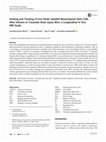
Stem cells transplantation has emerged as a promising alternative therapeutic due to its potency ... more Stem cells transplantation has emerged as a promising alternative therapeutic due to its potency at injury site. The need to monitor and non-invasively track the infused stem cells is a significant challenge in the development of regenerative medicine. Thus, in vivo tracking to monitor infused stem cells is especially vital. In this manuscript, we have described an effective in vitro labelling method of MSCs, a serial in vivo tracking of implanted stem cells at traumatic brain injury (TBI) site through 7 T magnetic resonance imaging (MRI). Proper homing of infused MSCs was carried out at different time points using histological analysis and Prussian blue staining. Longitudinal in vivo tracking of infused MSCs were performed up to 21 days in different groups through MRI using relaxometry technique. Results demonstrated that MSCs incubated with iron oxide-poly-L-lysine complex (IO-PLL) at a ratio of 50:1.5 μg/ml and a time period of 6 h was optimised to increase labelling efficiency. T2*-weighted images and relaxation study demonstrated a significant signal loss and effective decrease in transverse relaxation time on day-3 at injury site after systemic transplantation, revealed maximum number of stem cells homing to the lesion area. MRI results further correlate with histological and Prussian blue staining in different time periods. Decrease in negative signal and increase in relaxation times were observed after day-14, may indicate damage tissue replacement with healthy tissue. MSCs tracking with synthesized negative contrast agent represent a great advantage during both in vitro and in vivo analysis. The proposed absolute bias correction based relaxometry analysis could be extrapolated for stem cell tracking and therapies in various neurodegenerative diseases.
Stem Cells and Development, Jun 1, 2011
Homing and engraftment of hematopoietic stem/progenitor cells (HSPCs) in bone marrow is the major... more Homing and engraftment of hematopoietic stem/progenitor cells (HSPCs) in bone marrow is the major determining factor in success of hematopoietic stem cell transplantation. This is a complex, multistep process orchestrated by the coordinated interplay between adhesion molecules, cytokines, growth factors, and regulatory cofactors, many of which remain to be defined. Recent studies have highlighted the pivotal role of unique stromal-derived factor-1 (SDF-1)/CXCR4 signaling in the regulation of HSPC homing and subsequent engraftment. In addition, studies suggest that SDF-1/CXCR4 signaling acts as an essential survival-promoting factor of transplanted HSPCs as well as maintenance of quiescent HSCs in bone marrow niche. These pleiotropic effects exerted by SDF-1/CXCR4 axis make this unique signaling initiator very promising, not only for optimal hematopoietic reconstitution but also for the development of innovative approaches to achieve restoration, regeneration, or repair of other damaged tissues potentially amendable to reversal by stem cell transplantation. This goal can only be achieved when the role of SDF-1/CXCR4 axis in hematopoietic transplantation is clearly defined. Hence, this review presents current knowledge of the mechanisms through which SDF-1/CXCR4 signaling promotes restoration of hematopoiesis by regulating the homing and engraftment of HSPCs.
International Journal of Nanomedicine, Jun 1, 2021
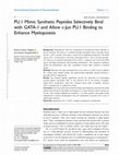
Background: Hematopoietic stem cells' commitment to myelopoiesis builds immunity to prevent infec... more Background: Hematopoietic stem cells' commitment to myelopoiesis builds immunity to prevent infection. This process is controlled through transcription factor, especially Purine rich box 1 (PU.1) protein, which plays a central role in regulating myelopoiesis. The β3/β4 region of PU.1 accommodates a coactivator transcription factor, c-Jun, to activate myelopoiesis. However, an erythroid transcription factor, GATA-1, competes with c-Jun for the β3/β4 region, abolishing myelopoiesis and promoting erythropoiesis. This competitive regulation decides the hematopoietic stem cells' commitment towards either erythroid or myeloid lineage. Methods: Therefore, this study investigated the in vitro and in vivo effect of novel synthetic PU.1 β3/β4 mimic peptide analogs and peptide-loaded hydrophilic poly(D,L-lactide-coglycolide) (PLGA) nanoparticles. Results: The designed peptides significantly increase the expression of corresponding myeloid markers, specifically CD33 in vitro. However, the in vivo delivery of peptideloaded PLGA nanoparticles, which have sustained release effect of peptides, increases 10.8% of granulocytes as compared to control. Conclusion: The observations showed that the fabricated nanoparticles protected the loaded peptides from the harsh intracellular environment for a longer duration without causing any toxicity. These findings highlight the possibility to use these peptides and peptide-loaded nanoparticles to increase hematopoietic stem cell commitment to myeloid cells in case of opportunistic infection.
Life Sciences, Dec 1, 2019
Endothelial cell damage is critical to understand since its presence in the entire body makes the... more Endothelial cell damage is critical to understand since its presence in the entire body makes the damage widespread instead of being localized. Being a major component of stem cell niche in bone marrow, deems it essential to gain knowledge of the damage to endothelium associated with bone marrow. Since radiation exposure has become common to numerous therapeutic modalities, its effects on bone marrow and its endothelial cells are crucial to understand. Material & methods: Microarray analysis was performed on irradiated human bone marrow endothelial cells (hBMECs) with and without prior treatment with radioprotectant amifostine to assess the effects of radiation on signalling pathways and the subsequent changes in pathways when treated with radioprotectant prior to radiation exposure. Key findings: It was seen that adhesion pathways that were usually inactivated under normal circumstances were stimulated post radiation. However, where in the case of radiation exposure, these adhesion pathways included leukocyte adhesion and migration; in the case of radioprotected conditions the pathways revolve around cellsubstrate adhesion and cell spreading. Genes like ROCK1, FLNA, RAC1, PRKCZ and MAP3K8 were seen to regulate the molecular switch between leukocyte-cell adhesion to cell-substrate adhesion. Significance: Our study demonstrated that irradiated endothelium supports leukocyte adhesion and migration but shifts to substrate adhesion dependent cell spreading under radioprotected conditions in order to repair the monolayer damage from the radiation. The genes responsible for the shift were identified and can be employed to manipulate cell adhesion characteristics for the treatment of diseases caused by radiation or inflammation.
Journal of Microencapsulation, Aug 18, 2018
A c c e p t e d M a n u s c r i p t Cell encapsulation potential of chitosan-alginate electrostat... more A c c e p t e d M a n u s c r i p t Cell encapsulation potential of chitosan-alginate electrostatic complex in preventing natural killer and CD8 + cell-mediated cytotoxicity: an in vitro experimental study
Journal of Nanoparticle Research, Jun 22, 2013
Single living Jurkat cells have been encapsulated through polyelectrolytes nanoparticles poly(all... more Single living Jurkat cells have been encapsulated through polyelectrolytes nanoparticles poly(allyl amine hydrochloride) size 15.6 nm and poly(styrene-co-sulfonic acid sodium salt) size 30.2 nm, through layer by layer coating of oppositely charged plasma membrane. Confocal microscopy and scanning electron microscopy results showed complete shielding of Jurkat cells, and no changes in cell surface morphology of encapsulated cells were observed. Cell viability was not affected after encapsulation and no toxicity was found. In vivo studies demonstrated no significant changes in hematological and biochemical parameters of blood serum at day 1 and 7 in mice. Histopathological analyses of liver and spleen tissues showed nontoxic nature of prepared formulations.
Experimental Cell Research, Feb 1, 2017
Mesenchymal stem cells (MSCs) are frequently used as a therapeutic, but reliable imaging techniqu... more Mesenchymal stem cells (MSCs) are frequently used as a therapeutic, but reliable imaging technique to longitudinally evaluate the engraftment of transplanted cells is inadequate. For magnetic resonance imaging (MRI), it is essential to understand the technical competence of in vitro stem cells labeling with iron oxide with regard to its relaxation behavior and significance of its biological expressions. The purpose of the study was to optimize the effective labeling of MSCs with high transverse relaxivity iron oxide contrast agent with protamine sulfate and also evaluate the biological effects (phenotype and function) of labeled MSCs. Our results demonstrated that 50:3 µg/ml of Fe-Pro complex containing 10% serum at an incubation time of 6 h were ideal for effective in vitro labeling. Relaxometry study demonstrated that almost an 8-fold increase in relaxation rate (R 2) was observed in labeled MSCs by comparing with unlabeled. Marginal alteration in Oct4 and CD146 genes, and phenotypic CD45 expressions were detected after labeling. T2-weighted images and histological analysis confirmed the homing of transplanted cells to the site of injury. The relaxometry based optimized labeling method of MSCs could be extrapolated for cellular MRI and may be useful in stem cell tracking in various pre-clinical and clinical studies.
Niche, Jan 6, 2016
Hematopoietic stem cells have dissimilar proliferative capacities; although some remain in quiesc... more Hematopoietic stem cells have dissimilar proliferative capacities; although some remain in quiescence, some consistently undergo multiple rounds of cell division stimulating hematopoiesis to maintain homeostasis and protect stem cell exhaustion. The stringent equilibrium between quiescence, self-renewal, proliferation, and differentiation is maintained by the operational selectiveness of intrinsic factors and extrinsic cues provided by the specialized microenvironment called the "niche" that provides homeness to hematopoietic stem cells (HSCs). The niche is defined by a heterogeneous identity involving the complex interaction of various other cells, cytokines, adhesion molecules, and extracellular matrix via several surface receptors and soluble factors, which work in paracrine coordination and protect stem cells from depletion while eliciting proliferation. The widespread investigations have shed light on the mechanistic underpinnings in niche, which coordinate the balance between relative hematopoietic quiescence and proliferation. In this paper, we discuss the recent findings concerning how these complex niche interactions facilitate the regulation of hematopoietic stem cells during homeostasis and further extend the discussion to their involvement in expansion and tumorigenesis, the two biological phenomena that are intimately associated with stem cells.
Journal of Stem Cells & Regenerative Medicine, Apr 30, 2013
Cell Adhesion & Migration, Jul 4, 2014
Homing and engraftment, a determining factor in hematopoietic stem cell transplantation success i... more Homing and engraftment, a determining factor in hematopoietic stem cell transplantation success is defined as a process through which hematopoietic stem/progenitor cells (HSPCs) lodge recipient bone marrow. SDF-1/CXCR4 axis acts as a principle regulator in homing and engraftment, however, CXCR4 signaling is dependent upon expression of CXCR4 and its ligand SDF-1, which is highly dynamic. Hence, present investigation was aimed to explore the potential of CXCR4 constitutive active mutants (CXCR4-CAMs) in overcoming the limitation of CXCR4 signaling and up-modulate its efficiency in homing and engraftment. Regulated transgene expression study of these mutants revealed their significantly enhanced cell adhesion efficiency to endothelium and extracellular matrix protein. This altogether indicates promising prospects of CXCR4-CAMs in research aimed to improve HSPCs engraftment efficiency.
Transplantation and Cellular Therapy, Oct 1, 2022
Journal of Cancer Therapy, 2016
The tyrosine kinase receptor III, c-Kit/stem cell factor receptor and its ligand, human stem cell... more The tyrosine kinase receptor III, c-Kit/stem cell factor receptor and its ligand, human stem cell factor (huSCF) are the predominant regulator of mitogenesis in the hematopoietic stem and progenitor cells. However, gain-of-function mutations alter c-Kit auto-regulatory mechanisms to aberrant c-Kit signaling, leading to the onset or progression of cancerous transformations. The most common mutation of c-Kit is the substitution of aspartic acid residue in position 816 to valine (D816V), which is majorly responsible for its ligand-independent constitutive activation, and is implicated in hematopoietic malignancies. Currently, molecular targeted therapy is increasingly becoming a hot spot due to its specificity and low toxicity. As the molecular mechanisms responsible for D816V-c-Kit mediated tumorogenicity are largely unknown, in this study, we aimed to investigate the D816V-c-Kit signaling mediated downstream molecular targets. Specifically, we created c-Kit active mutant form D816V and performed inducible gene expression of mutant D816V-c-Kit in monomyelocytic cell line U937. Mutant D816V-c-Kit expressing cells revealed significantly enhanced cellular mitogenic activity compared to wild-type c-Kit expressing cells independent of huSCF. To examine the molecular targets regulating tumorogenic proliferation, we evaluated the consequences of mutant D816V-c-Kit expression on downstream gene expression profile by high throughput microarray technology. The levels of some of the relevant genes (PIK3CB, eIF4B, PRKCDBP, MOAP1) were validated by quantitative polymerase chain reaction. SLA, STAT5B, MAP3K2 and MAPK14 emerged as important downstream molecular targets of mutant D816V-c-Kit. Further, by dissecting the signaling pathways, we also demonstrated that the D816Vc-Kit mediated hematopoietic cell proliferation is dependent on molecular target p38 MAP kinase.
European Journal of Neuroscience, May 12, 2021
Cellular transplantation of stem cells can be a beneficial treatment approach for neurodegenerati... more Cellular transplantation of stem cells can be a beneficial treatment approach for neurodegenerative diseases such as traumatic brain injury (TBI). In this study, we investigated the proliferation and differentiation potential of infused mesenchymal stem cells (MSCs) after localisation at the injury site. We evaluated the appropriate homing of infused MSCs through immunohistochemistry, followed by Y‐chromosome–specific polymerase chain reaction and fluorescent in situ hybridisation analyses. The proliferation and differentiation of infused MSCs were analysed using exogenous cell tracer 5′‐bromo‐2′‐deoxyuridine (BrdU) labelling and neuronal specific markers, respectively. Structural and functional recovery in TBI mice were examined by performing magnetic resonance imaging and different behavioural assessments, respectively. Results demonstrated a significantly high number of BrdU‐positive cells in the lesion region in the MSC‐infused group compared with control and TBI groups. Infused MSCs were well differentiated into neural‐like cells and expressed significantly more neural markers (neuronal nuclear antigen [NeuN], microtubule‐associated protein 2 [MAP2] and glial fibrillary acid protein [GFAP]). Improved tissue abnormalities as well as functional behaviours were observed in MSC‐infused TBI mice, implying the substantial proliferation and differentiation of infused MSCs. Our findings support the neuroprotective response and efficacy of MSCs after transplantation in TBI mice, and MSCs may serve as potential therapeutic candidates in regenerative medicine.


Uploads
Papers by Gurudutta Gangenahalli